Endometeriosis
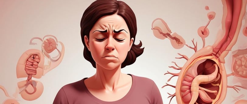
Endometriosis is a condition that can affect females. It happens when tissue that is similar to that of endometrial tissue grows outside the uterus.
DISEASES & DISORDERS
Endometriosis
Traditionally, endometriosis is defined as the presence of endometrial glands and tissue outside the usual location of the uterus and its muscles. However, some definitions require that this misplaced tissue must be able to function like normal endometrial tissue. Initially, endometriosis was categorized into two types: endometriosis interna and endometriosis externa. Endometriosis interna, which is now known as adenomyosis, involves the benign growth of endometrial tissue into the muscular wall of the uterus (myometrium). On the other hand, endometriosis externa refers to the presence of endometrial tissue in locations other than the uterus, such as near the uterus (like the fallopian tubes and ovaries) or even in more distant sites like the brain.
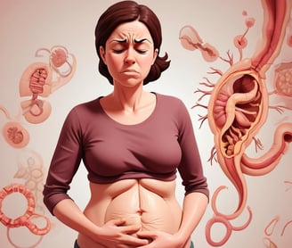
Symptoms of endometriosis include :
Endometriosis often causes painful cramps, similar to menstrual cramps, along with persistent lower back and pelvic pain. Women with this condition may experience periods that last longer than a week, heavy menstrual bleeding, and bowel or urinary issues like pain, diarrhea, constipation, or bloating. They might also notice blood in their stool or urine, feel nauseous or vomit, experience fatigue, have pain during sex, and occasionally have spotting or bleeding between periods. Interestingly, while pain is the most common symptom of endometriosis, its severity doesn't always match how extensive the disease is.
Endometriosis can lead to infertility due to factors like distorted pelvic anatomy, impaired egg quality, inflammation, hormonal imbalances, and treatment effects. While not all women with endometriosis face infertility, those who do have options like ovarian stimulation, IUI, IVF, or surgery to improve their chances of pregnancy.
Why and how does it occur?
This disease was first described by Daniel Shroen in 1690 in the work “Disputatio Inauguralis Medica de Ulceribus Ulceri”. The symptoms of this disease were presented by Arthur Duff in 1769 [1].
The first appearances in the literature regarding the pathogenesis of endometriosis appeared in the second half of the nineteenth century. This condition was described by Karl von Rokitansky in 1860, who defined it as the presence of an active endometrium outside the uterine cavity [2].
In 1882, Von Recklinghausen suggested the name adenomyoma and by the end of the nineteenth century, several more authors had described this disease.
In 1908, a monograph was published by T.S. Cullen on adenomyosis [3].
Cullen was the first to describe the two main symptoms of adenomyosis: prolonged menstrual bleeding and severe pain. He believed that endometrial tissue came from the remains of Müller’s ducts [3,4].
In 1870, the German anatomist, physiologist and pathologist Heinrich Wilhelm Waldeyer was the first to put forward the theory of metaplasia.
One of the outstanding doctors of the nineteenth century, Iwanhofen in 1898 was the author of the thesis that endometrial tissue arises from metaplasia of the peritoneal epithelium.
In 1927, J.A. Sampson was the first to introduce the term “endometriosis” into medical nomenclature. According to the researcher, the cause of the disease is “retrogradea menstruation” or retrograde transport of menstrual blood with the consequent implantation of exfoliated endometrial mucosa cells within the peritoneal cavity.
Despite the passage of such a long period of time and much scientific research, Sampson’s theory is still dominant among other hypotheses regarding the etiopathogenesis of endometriosis. It has still not been possible to fully explain why the retrograde transport of menstrual blood, which occurs in nearly 90% of women of childbearing age, only leads to the survival of endometrial tissue outside the uterine cavity in a minority of women [7].
Risk Factors for Endometriosis Include:
Early menarche—epidemiological studies analyzing the cycle of women with endometriosis have shown that the early first cycle (before the age of 11) is associated with the risk of endometriosis [43,44,45,46],
Shorter than 27-day genital cycles, genital defects, including hymen overgrowth or narrowing of the cervical canal [47]. The risk of endometriosis is increased in women with short cycles, i.e., lasting less than 27 days, but is unrelated to the number of bleeding days and the volume of menstruation [48],
Low BMI,
Small number of births,
Caucasian race,
Age 25–29,
Daily consumption of alcohol in the amount of at least 10 g per day,
Endometriosis is more often diagnosed in infertile women who are active smokers and whose body mass index (BMI) is normal or low [49].
There are several types of endometriosis:
Ovarian endometriosis—occurs in the form of superficial lesions and as endometrial cysts,
Peritoneal—can occur in various forms: white raids on the peritoneum, peritoneal defects, red, brown, black-blue and black foci, colorless bright vesicles and focal dilated blood vessels and petechiae,
Deep infiltrating endometriosis—DIE,
Endometriosis of other locations.
Endometriosis comes in different types:
1. Peritoneal Endometriosis: This type can be either intraperitoneal or sub-peritoneal. It's found in 15-50% of women with endometriosis and is often detected during laparoscopic surgery.
2. Ovarian Endometriosis (Chocolate cysts) : Seen in 2-10% of reproductive-age women and 50% of those with infertility issues, it's one of the most common forms of endometriosis.
3. Deep Infiltrating Endometriosis (DIE):** This type involves endometrial changes that reach deep into the pelvis, affecting organs like the bladder, ureters, large intestine, ligaments, or vagina. The exact causes of DIE are not well understood.
Diagnosis:
Endometriosis is diagnosed using various methods:
1. Ultrasound Examination: This is the basic diagnostic test for endometriosis. It helps identify endometrial cysts in the ovaries and reproductive organ defects that can cause menstrual blood to flow backward into the abdomen.
2. Additional Tests: For cases where endometriosis affects the urinary bladder or large intestine, other tests like cystoscopy, colonorectoscopy, and transrectal ultrasound may be needed.
3. Deep Infiltrating Endometriosis: For deeper cases, Rectal Water Contrast Transvaginal Sonography (RWC TVS) can be helpful. This method uses water contrast to detect endometriosis in the intestinal area and track its progression.
4. Gold Standard: The most reliable way to diagnose endometriosis is through laparoscopic surgery, followed by a histopathological examination to confirm the findings.
Treatment:
The goal of pharmacological treatment is to reduce or eliminate pain, inhibit further development and regression of endometrial foci and restore fertility.
Sources:
Smolarz, B., Szyłło, K., & Romanowicz, H. (2021). Endometriosis: Epidemiology, Classification, Pathogenesis, Treatment and Genetics (Review of Literature). International Journal of Molecular Sciences, 22(19), 10554. https://doi.org/10.3390/ijms221910554
Jubanyik, K. J., & Comite, F. (1997). EXTRAPELVIC ENDOMETRIOSIS. Obstetrics and Gynecology Clinics of North America, 24(2), 411–440. https://doi.org/10.1016/S0889-8545(05)70311-9